2026 Class 7 Science · English Version · Chapter 2 · Chapter companion part
Chapter 2A: Inside plant and animal cells
Draw a labelled cell, connect each structure to its job and compare plant and animal examples using evidence.
Class 7 English Version: part A of the companion for chapter 2, Cellular Organisation of Plants and Animals. Eight teaching sections, 30 written tasks and a notebook challenge. Selected companion coverage, not complete textbook coverage; practical and independent review remain.
A cell is a basic structural and functional unit of living organisms. Some organisms are one cell; multicellular organisms have specialised cells. The textbook combines organelles into typical cell drawings. Such a drawing is a study model, not a microscope photograph of one cell containing every possible structure. A plant cell need not be a perfect rectangle and an animal cell need not be a perfect circle. Compare structures rather than outline alone.
Wall, membrane and cytoplasm
In a plant cell, the cellulose-rich wall is outside the plasma membrane. The wall supports and protects; the selectively permeable membrane regulates exchange with the surroundings. It is one membrane made from a phospholipid bilayer, not two separate membranes. Animal cells have a membrane but no cellulose wall. Cytoplasm is the material inside the membrane outside the nucleus, including fluid and organelles. Protoplasm is an older collective term for living cell contents; it is not a separate organelle.
Plastids and the central vacuole
Chloroplasts contain chlorophyll and carry out photosynthesis. Their envelope has an outer and inner membrane; internal thylakoid membranes form stacks called grana, with fluid stroma around them. Chromoplasts contain coloured pigments; colourless leucoplasts can store materials such as starch. Not every plant cell has chloroplasts: an underground root is an important counterexample. A mature plant cell commonly has a large sap-filled central vacuole. Its membrane is the tonoplast. Vacuoles support storage and water balance, while animal cells can have smaller membrane-bound compartments.
Mitochondria transfer energy
Both plant and animal cells can contain mitochondria. Their outer membrane surrounds an inner membrane folded into cristae, with matrix inside. During aerobic respiration, chemical energy from nutrients is transferred into ATP, a molecule used to power cell work. Energy is transformed, not created. Some ATP-making steps occur outside mitochondria, so the nickname powerhouse is a useful reminder rather than a complete explanation. The small inner-membrane particles called oxisomes in the source are associated with ATP synthase; they are not extra nuclei.
Making, processing and moving proteins
Ribosomes build proteins from amino acids. They can be free or attached to rough endoplasmic reticulum, a membrane network. Smooth endoplasmic reticulum lacks attached ribosomes and has roles including lipid production. The Golgi apparatus modifies, sorts and packages materials arriving in vesicles; it does not replace ribosomes as the site of protein synthesis. Lysosomes contain digestive enzymes inside a single membrane. Plant vacuoles can perform related breakdown roles. A typical animal centrosome contains paired centrioles that help organise microtubules; most flowering-plant cells lack centrioles but can still divide.
Read the nucleus carefully
The nuclear envelope has two membranes and pores that regulate exchange with the cytoplasm. Nucleoplasm is the internal fluid environment. The nucleolus helps assemble ribosomal subunits. Chromatin is DNA associated with proteins; it becomes more condensed as a cell prepares to divide. Chromosomes are not made by randomly breaking a nuclear net into pieces. A nucleus directs many activities through genetic information, but not every specialised cell retains one. A typical-cell diagram describes an example, not an exceptionless rule.
Compare with a structured table
Compare the same features in the same order: membrane, wall, nucleus, mitochondria, plastids and vacuole. Use present, absent or varies with cell type, and give a reason. A leaf cell can have chloroplasts and a large vacuole while an underground root cell has no green chloroplasts. Animal and plant examples both need membranes and ribosomes. A table makes similarities as visible as differences; do not choose only features that make one group seem better.
Use a microscope and a calculation honestly
A labelled study diagram can show details beyond a classroom light microscope. Optical magnification is eyepiece magnification multiplied by objective magnification: a 10× eyepiece and 40× objective give 400×. This enlarges the image; it does not make the real cell larger or guarantee that two nearby features can be distinguished. If image size and real size are known, use the same units before dividing. Draw only observed details in an observation record, and keep model labels in a separate annotated drawing. Use supplied samples or teacher-supervised equipment, not self-collected body material.
Worked example
A 10× eyepiece with a 40× objective gives 10 × 40 = 400×. For a separate illustrative drawing, 30 mm ÷ 0.05 mm = 600×. The first calculation uses lens settings; the second compares measured lengths. Neither number establishes the detail an instrument can resolve.
BoundaryWall outside membrane in plant model
Protein synthesisRibosome
ProcessingER and Golgi pathway
Genetic materialChromatin in nucleus
Energy transferMitochondrion and ATP
Compare structures using the same feature and function, including exceptions.
Read the labelled model and connect each structure to its job.
Show a comparison or calculation with the relevant evidence and units.
Check the model answer, correct one misconception and try the notebook task.
Source wording and corrections
Printed pages 13–16 contain compressed or misleading wording. The wall is outside the plasma membrane. A lysosome has one membrane made from a lipid bilayer. Chromatin condenses rather than breaking into chromosome pieces. Fungi are not lower plants. Mitochondria do not create all energy. These corrected explanations are the answer basis here.
A cautious colour explanation
The source shows a carrot without a colour photograph and calls it red. Chromoplast pigments account for many root colours; orange carrots commonly contain beta-carotene, while red varieties can differ. Do not infer one exact pigment from a black-and-white drawing. Covered grass can become pale as chlorophyll is lost or not maintained in darkness; avoid claiming every plastid always converts in the same way.
Compare the same cell features
Read across one row to compare the same feature. Then read down a column to describe a cell. These are typical examples, not counts from an experiment.
Compare the same cell features
Feature
Plant example
Animal example
Plasma membrane
Present, inside wall
Present; outer cell boundary
Cellulose wall
Present
Absent
Nucleus
Present in this typical living cell
Present in this typical cell
Mitochondria
Present
Present
Chloroplast
Present in photosynthetic cells; not all roots
Absent
Vacuole
Often a large central compartment in mature cells
Smaller compartments can occur
Ribosomes
Present; free or on rough ER
Present; free or on rough ER
Worked reading: mitochondria are present in both examples, so they are a similarity. Chloroplast presence depends on which plant cell is selected.
Magnification: two different calculations
Magnification describes how many times larger an image length is than the actual length. It has no length unit because the equal units cancel.
M = I ÷ A | optical M = eyepiece × objective
M
Magnification, a ratio with no length unit.
I
Measured image length.
A
Actual object length, expressed in the same units as I.
Length example: image I = 30 mm, actual A = 0.05 mm; use matching units.
M = 30 mm ÷ 0.05 mm = 600. The image length is 600 times the actual length.
Check: 0.05 mm × 600 = 30 mm. The real cell has not grown.
Original extension to the chapter’s microscope study, not a copied textbook calculation. The lengths are invented teaching values. None of the study plates has this measured scale. Higher magnification alone does not guarantee that more detail can be resolved.
Read the scientific images
Match each letter to its structure and function. These original study diagrams are simplified models, not microscope observations.
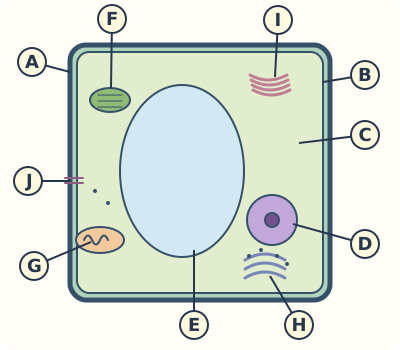
Read a typical plant-cell model
Trace the outer wall, then the membrane, then the cytoplasm. Explain why the vacuole boundary is a different membrane from the cell boundary.
A · Cell wall
Cellulose-rich support outside the plasma membrane; not the selectively permeable membrane.
B · Plasma membrane
The boundary just inside the wall regulates exchange. It is one phospholipid-bilayer membrane.
C · Cytoplasm
Fluid and organelles outside the nucleus, inside the cell membrane.
D · Nucleus
The genetic-information compartment; its detailed structures are shown in the separate nucleus plate.
E · Vacuole and tonoplast
The large central compartment contains cell sap; the line around it is its own membrane, the tonoplast.
F · Chloroplast
A plastid for photosynthesis in this green-cell example; many root cells lack it.
G · Mitochondrion
Transfers nutrient energy into ATP through stages of aerobic respiration; plants need mitochondria too.
Modifies, sorts and packages cellular materials; it does not replace ribosomes.
J · Plasmodesma
A channel crossing the wall connects adjacent plant cells; the neighbouring cell is outside this simplified view.
Original typical-cell schematic, not to scale or a microscope photograph. The source combines structures from different cells. The membrane belongs inside the wall.
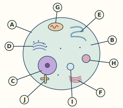
Read a typical animal-cell model
Compare this model with the plant cell using the same row headings. Include shared structures as well as differences.
A · Plasma membrane
The outer cell boundary; there is no cellulose wall in this animal model.
B · Cytoplasm
The region around the organelles and outside the nucleus.
C · Nucleus
Contains DNA with proteins as chromatin; envelope, pores and nucleolus have separate roles.
D · Rough ER and ribosomes
Ribosomes on the network build proteins entering this pathway. Free ribosomes also occur.
E · Smooth ER
A membrane network without attached ribosomes; roles include lipid production.
F · Golgi apparatus
Modifies and packages incoming materials into vesicles.
G · Mitochondrion
Has an inner and outer membrane and participates in aerobic ATP production.
H · Lysosome
A single-membrane compartment containing digestive enzymes; one lipid bilayer is one membrane.
I · Vesicle / small compartment
A membrane-bound sac used in transport or storage; it is not a giant central plant vacuole.
J · Centrosome / paired centrioles
Helps organise microtubules in this typical animal model. Most flowering-plant cells lack these centrioles but can divide.
Original composite schematic, not a claim that every animal cell has this exact shape or every labelled organelle. Detail visible in this model may not be resolvable with a school light microscope.
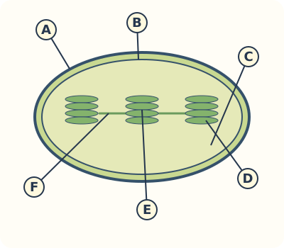
A chloroplast has several membrane systems
Identify an envelope, one flattened compartment and a stack. Different sizes of membrane structure should not all receive the same label.
A · Outer envelope membrane
The outer of two membranes surrounding the chloroplast.
B · Inner envelope membrane
The inner envelope encloses the stroma. It is distinct from the internal thylakoids.
C · Stroma / matrix
Fluid around the thylakoid system; the source also labels matrix. Do not confuse it with mitochondrial matrix.
D · Thylakoid
One flattened membrane compartment. Chlorophyll is associated with thylakoid membranes.
E · Granum / grana
A stack of thylakoids is a granum; grana is plural. The source’s granum lamella refers to membranes in this stack.
F · Stroma lamella
A membrane connection between stacks; it is not a third envelope surrounding the chloroplast.
Original schematic, not to scale. This detailed model is not what every green blob under a light microscope reveals. The outer/inner envelope and thylakoid system must stay distinct.
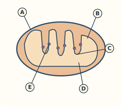
Mitochondrial folds and energy transfer
Connect folds to membrane area and ATP production. Explain that nutrients supply chemical energy which is transferred rather than newly created.
A · Outer membrane
The outer boundary of this double-membrane organelle.
B · Inner membrane
The membrane enclosing the matrix; it folds inward.
C · Crista / cristae
One fold is a crista, multiple folds are cristae; folds increase inner-membrane area.
D · Matrix
The internal compartment. Different respiration reactions occur in different locations.
E · ATP-synthase particles
Inner-membrane complexes associated with ATP formation; the source calls particles oxisomes. They are not extra chromosomes or nuclei.
Original schematic, not an electron micrograph or an exact particle count. Mitochondria are not the site of every energy-releasing reaction in a cell.
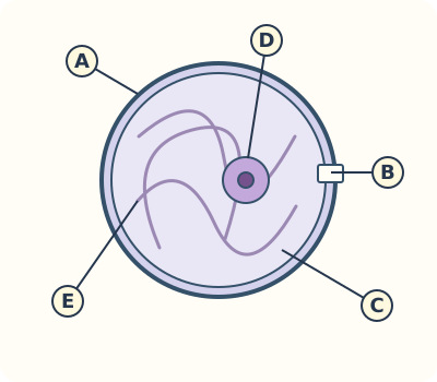
Five nuclear labels, five different meanings
Use the leader lines to distinguish boundary, opening, fluid, assembly region and genetic material. Redraw the model with functions beside the labels.
A · Nuclear envelope
Two membranes separate the nucleus from the cytoplasm.
B · Nuclear pore
A regulated passage through the envelope; it is not an unregulated tear.
C · Nucleoplasm
The internal fluid environment around nuclear structures.
D · Nucleolus
A region involved in ribosomal-subunit assembly; not a second nucleus.
E · Chromatin
DNA with associated proteins. It condenses into more visible chromosome structures as a cell prepares to divide.
Original schematic. Source wording about a nuclear net breaking into pieces is corrected to chromatin condensation; random DNA fragmentation does not form healthy chromosomes.
Your written practice
Show your working on paper and enter your answer here. If it does not match, compare the model and try again.
Show model answer and explanation
cell wall
The wall supports the cell; the membrane is inside it.
Show model answer and explanation
cell membrane
The plasma membrane is selectively permeable.
Show model answer and explanation
ribosomes
Ribosomes join amino acids in protein synthesis.
Show model answer and explanation
Golgi apparatus
The Golgi receives materials, modifies them and packages them.
Show model answer and explanation
cristae
Cristae increase the inner-membrane area used in ATP production.
Show model answer and explanation
stroma
Do not confuse chloroplast stroma with mitochondrial matrix.
Show model answer and explanation
chloroplast
Chloroplasts perform photosynthesis.
Show model answer and explanation
tonoplast
The tonoplast is distinct from the outer cell membrane.
Show model answer and explanation
nucleolus
It is different from the whole nucleus.
Show model answer and explanation
400
Multiply 10 by 40; do not add them.
Write in your own words. A guardian or teacher should review this response.
Show model answer and explanation
Put the wall on the outside, a separate membrane just inside it, and cytoplasm inside the membrane around the organelles. State that the drawing is a model.
Check the position of each leader line, not only the spelling.
Write in your own words. A guardian or teacher should review this response.
Show model answer and explanation
A photosynthetic leaf cell can have chloroplasts. An underground root cell normally lacks green chloroplasts. The animal cell has no chloroplasts. All three can contain mitochondria.
A plant classification alone does not prove chloroplasts are present.
Write in your own words. A guardian or teacher should review this response.
Show model answer and explanation
They transfer chemical energy from nutrients into ATP during aerobic respiration. ATP powers cell activities; energy changes form rather than being created.
Include ATP and energy transfer, and acknowledge the nickname is simplified.
Write in your own words. A guardian or teacher should review this response.
Show model answer and explanation
A ribosome on rough ER makes the protein. It enters the ER pathway, travels in a vesicle to the Golgi, and is processed and packaged into a transport vesicle.
The Golgi does not take the ribosome’s protein-building role.
Write in your own words. A guardian or teacher should review this response.
Show model answer and explanation
A bilayer is the two lipid layers within one membrane. A lysosome has a single surrounding membrane; a nuclear envelope has two membranes.
Distinguish a lipid layer from a whole membrane.
Write in your own words. A guardian or teacher should review this response.
Show model answer and explanation
The envelope separates nucleus from cytoplasm and has pores. The nucleolus helps assemble ribosomal subunits. Chromatin is DNA with proteins carrying genetic information.
A location name alone is not a function.
Write in your own words. A guardian or teacher should review this response.
Show model answer and explanation
Chromatin becomes more condensed into distinguishable chromosomes. This is organised packing of DNA and proteins, not random fragmentation.
Use condensation rather than breakage.
Write in your own words. A guardian or teacher should review this response.
Show model answer and explanation
Rubbing or breaking potato tissue can release stored starch grains from cells. A grain in a drop is not itself proof of an intact cell, a nucleus or a complete plastid.
Separate the observed particle from the structure it came from.
Write in your own words. A guardian or teacher should review this response.
Show model answer and explanation
Use the same six row headings for both: membrane, wall, nucleus, mitochondria, plastids and vacuole. Mark typical features and relevant exceptions, and label both drawings as models.
Include shared structures and a genuine variation, not only differences.
Write in your own words. A guardian or teacher should review this response.
Show model answer and explanation
Magnification = 30 mm ÷ 0.05 mm = 600. The image length is 600 times the real length; the cell itself has not grown.
Use the same length units so they cancel. This is illustrative measurement data, not a scale for the study plates.
Show model answer and explanation
E
E identifies the compartment and its membrane.
Show model answer and explanation
J
J marks the paired-centrioles example.
Show model answer and explanation
D
E is the granum stack; D is one compartment.
Show model answer and explanation
D
D is inside the inner membrane.
Show model answer and explanation
E
E points to DNA-associated threads.
Write in your own words. A guardian or teacher should review this response.
Show model answer and explanation
A is the outside wall and B the membrane just inside it. Redraw the boundaries in that order and connect support to wall and selective exchange to membrane.
Use both position and function.
Write in your own words. A guardian or teacher should review this response.
Show model answer and explanation
D is one flattened thylakoid, E is a stack called a granum, and F links stacks through the stroma. They are parts of the internal membrane system rather than extra envelope membranes.
Differentiate a component, a stack and a connection.
Write in your own words. A guardian or teacher should review this response.
Show model answer and explanation
The nuclear envelope has two membranes with pores; a lysosome has one surrounding membrane. Each membrane itself is a phospholipid bilayer.
Two lipid layers do not mean two membranes.
Write in your own words. A guardian or teacher should review this response.
Show model answer and explanation
It is an explanatory schematic incorporating information beyond a routine classroom light-microscope view. Its labels are model information, not reported observations.
Separate a useful model from measured resolution.
Write in your own words. A guardian or teacher should review this response.
Show model answer and explanation
Keep the wall, membrane, cytoplasm, nucleus and appropriate organelles, but do not automatically copy green chloroplasts from the leaf-like model. Storage plastids may be relevant.
Explain the choice rather than colouring every plant cell green.
Your science notebook challenge
Create a two-page cell atlas with plant and animal models, a comparison table and one magnification calculation.
Review with a guardian or teacher
Use accurate leader lines for the wall, membrane, nucleus, mitochondria and other studied organelles.
Link each label to a job and state one typical-cell exception.
Show calculation steps and units; separate a model from an observation.
Keep your work on paper or in a local file. This page does not upload it or automatically grade the project.
Chapter companion and further study
This original Kidemy practice follows the topic order of 2026 Class 7 Science · English Version, chapter 2, printed pages 12–24. It does not replace the complete chapter or its practical teaching.