2026 Class 7 Science · English Version · Chapter 1 · Chapter companion part
Chapter 1A: Microbes, viruses and bacterial cells
Compare microbes using cells, nuclei, shape and evidence rather than treating every small organism as the same.
Class 7 English Version: part A of the companion for chapter 1, Lower Organisms. Eight teaching sections, 30 written tasks and a notebook challenge. Selected companion coverage, not complete textbook coverage; practical and independent review remain.
Microorganisms are organisms too small to study clearly with unaided eyes. Some fungi and algae are large enough to see. The chapter title Lower Organisms is a school grouping, not a claim that small life is unimportant or less successful. Microbiology also studies viruses, which are acellular: they are not made of cells.
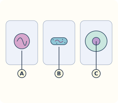
Compare three structural descriptions
The source diagram uses Akaryota for acellular agents, Prokaryota for cells without a membrane-bound nucleus, and Eukaryota for cells with such a nucleus. These are the book’s comparison labels, not three universally accepted modern kingdoms. Bacteria are prokaryotic; Amoeba, fungi and algae in these examples are eukaryotic. A small organism is not automatically prokaryotic.
What a virus particle contains
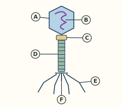
A virus carries genetic material, DNA or RNA, in a protein coat called a capsid; some viruses also have an envelope. A particle is not a tiny complete cell: it has no ribosomes or independent cellular metabolism. Viruses depend on suitable living host cells to make more virus particles. A bacteriophage is a virus that infects bacteria; its head-and-tail drawing is not the shape of every virus.
What a bacterial cell contains
A bacterium is a single cell with a membrane, cytoplasm, ribosomes and genetic material, but no membrane-bound nucleus. Many have a cell wall. Some bacteria can photosynthesise, so the source description non-green is not a rule for all bacteria. Bacteria can reproduce by binary fission, in which one cell divides into two; viruses cannot do this on their own.
Use shape words precisely
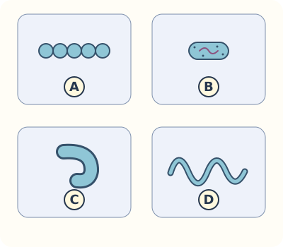
Coccus describes a roughly spherical bacterium; bacillus describes a rod; a vibrio is curved like a comma; a spirillum is spiral-shaped. The book uses Comma as the third shape label. A shape is a descriptive clue, not enough to identify a species or diagnose a disease. A coloured sketch is a model, not an observation through a microscope.
An Amoeba is a whole cell
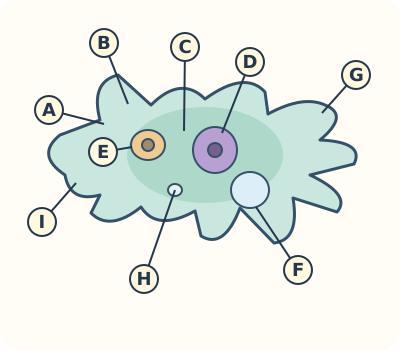
Amoeba has a cell membrane, cytoplasm and a nucleus. Temporary extensions called pseudopodia help it move and surround food. Food vacuoles hold engulfed material; a contractile vacuole helps remove excess water in a freshwater example. These are different jobs. Its changing outline does not mean it lacks a cell boundary or organised internal structures.
What images can support
A microscope magnifies an image; useful detail also depends on its resolving ability, meaning whether close features can be distinguished separately. A routine school light microscope does not show every detail of a virus or bacterium. Use supplied images, their labels and scale bars. State when information is not visible instead of filling a drawing with imagined observations.
Build a reasoned comparison
Compare an acellular virus, a bacterial cell and Amoeba in columns: cell present, membrane-bound nucleus, and how more individuals or particles are produced. A virus needs a host cell; a bacterium can divide; an Amoeba is a nucleated cell. Neither small size nor harmfulness alone separates these groups. Leave disease identification to qualified professionals, not a shape-matching game.
Worked example
A supplied particle has genetic material inside a protein coat and uses a host cell: it fits the virus model. A second example has cytoplasm, ribosomes and DNA but no nuclear membrane: it fits a bacterial cell. These descriptions do not identify a disease.
VirusAcellular; host-dependent replication
BacteriumCell; no membrane-bound nucleus
AmoebaCell with a nucleus
Coccus / bacillusSpherical / rod-shaped
Vibrio / spirillumComma-shaped / spiral
Structural comparison: size alone does not tell whether a nucleus is present.
Identify the structure, evidence or route named in the question.
Link each feature to its function; include a labelled sketch where useful.
Compare with the model, explain a correction and try the independent task.
Book wording and correction
Printed page 2 describes microorganisms as primitive organisms and then includes nucleated microbes. Use its later structural comparison: Amoeba and fungi have nuclei; bacteria do not have a membrane-bound nucleus. Microscopic does not mean primitive.
Try another comparison
A supplied yeast cell has a nucleus and ribosomes. It belongs with eukaryotic cells even though it is small; the presence of a nucleus is more useful than size.
Read the scientific images
Match each letter to its structure and function. These original study diagrams are simplified models, not microscope observations.
Compare cell organisation
Compare the presence of a cell and a nuclear compartment, rather than size or harmfulness.
A · Acellular
A virus particle has genetic material and a coat but is not a cell.
B · Prokaryotic cell
The bacterial model has DNA and ribosomes but no membrane-bound nucleus.
C · Eukaryotic cell
A nucleus encloses genetic material in the illustrated nucleated cell.
The book uses Akaryota, Prokaryota and Eukaryota as comparison groups. This is not a universal modern three-kingdom classification.
A T2-type bacteriophage model
Follow the structure from head to attachment apparatus. A phage infects bacteria; this shape does not represent every virus.
A · Head / capsid
The protein coat encloses the genetic material. Its geometric outline is a model.
B · DNA
The T2-type example contains DNA. Other viruses may contain RNA instead.
C · Collar
A neck region connects the head to the tail assembly.
D · Tail sheath
The contractile sheath surrounds the tail tube in this model; it is not a leg or a cell membrane.
E · Tail fibres
Fibres help recognise and attach to a suitable bacterial host.
F · Baseplate
The tail-end assembly contributes to attachment and delivery through the tail tube.
Original schematic, not to scale or a microscope image. The source labels head, DNA, collar, tail and tail fibre; baseplate and sheath/tube distinction add explanation.
Four bacterial shape models
Name the visible shape and compare it with another. A shape by itself does not identify species or establish disease.
A · Coccus
Roughly spherical. Cocci is the plural form; cells may occur singly, in pairs, chains or other groups. The plate shows a chain.
B · Bacillus
Rod-shaped. Bacilli is the plural shape word.
C · Vibrio / comma
Curved like a comma; the source calls this Comma.
D · Spirillum
A spiral outline; spirilla is the plural.
Original schematic at a common display size, not actual cell-size measurements. Source figure 1.4 does not clearly match its comma-shaped description; this plate shows a curved vibrio. A thin flagellum is a separate structure, not the bacterial cell body. Colours are study labels.
A freshwater Amoeba model
Trace how the cell extends around food, then explain why food processing and excess-water removal are different functions.
A · Membrane / plasmalemma
The cell boundary controls exchange; a changing outline still has a boundary.
B · Ectoplasm / plasma gel
The clearer outer cytoplasm is represented as a gel-like region, not a separate cell wall.
C · Endoplasm / plasma sol
The more fluid inner cytoplasm contains structures and can flow during movement.
D · Nucleus
Contains genetic material within a membrane-bound compartment.
E · Food vacuole
Contains engulfed material; it has a different role from the contractile vacuole.
F · Contractile vacuole
Collects and expels excess water in this freshwater model.
G · Pseudopodium
Temporary extension used in movement and food capture; not a permanent leg.
H · Water-filled space
Represents a small vacuolar space labelled water vacuole in the source; a circle alone does not establish its function.
I · Uroid / rear region
The trailing end in a moving Amoeba. The source spells its label Uriod; this is not a separate organ.
Original schematic, not to scale. Outer/inner cytoplasm names describe regions and physical state, not extra membranes. All nine lettered details have a text guide.
Your written practice
Show your working on paper and enter your answer here. If it does not match, compare the model and try again.
Show model answer and explanation
acellular
Acellular distinguishes a virus particle from a bacterial cell.
Show model answer and explanation
capsid
Some viruses also have an envelope; the capsid is the protein coat.
Show model answer and explanation
bacteriophage
A bacteriophage infects a bacterium.
Show model answer and explanation
prokaryotic
Bacteria have genetic material without a membrane-bound nucleus.
Show model answer and explanation
eukaryotic
Amoeba has a membrane-bound nucleus.
Show model answer and explanation
coccus
Coccus names the spherical shape, not a disease.
Show model answer and explanation
bacillus
Bacillus is the singular shape word.
Show model answer and explanation
vibrio
The chapter labels this shape Comma.
Show model answer and explanation
pseudopodia
Pseudopodia are extensions of the cell.
Show model answer and explanation
binary fission
This cell-division process is different from viral replication in a host.
Write in your own words. A guardian or teacher should review this response.
Show model answer and explanation
Amoeba and yeast are microscopic eukaryotes with nuclei. A bacterium is prokaryotic. Size alone does not establish cell organisation.
Give a counterexample and the relevant structural distinction.
Write in your own words. A guardian or teacher should review this response.
Show model answer and explanation
A bacterium is a cell with ribosomes and can divide under suitable conditions. A virus particle is acellular and uses a living host cell to produce more particles.
Do not claim that a virus divides like a bacterium.
Write in your own words. A guardian or teacher should review this response.
Show model answer and explanation
Use it to compare acellular agents, non-nucleated cells and nucleated cells. Modern classification uses further evidence and does not recognise these three labels as one universal kingdom scheme.
Separate the source’s classroom model from a universal classification claim.
Write in your own words. A guardian or teacher should review this response.
Show model answer and explanation
Keep the genetic material but remove the membrane-bound nucleus label; bacteria do not have that nuclear compartment.
Lacking a nucleus does not mean lacking DNA.
Write in your own words. A guardian or teacher should review this response.
Show model answer and explanation
Use spirillum for a spiral shape; coccus describes a roughly spherical cell. Shape supports a descriptive label, not a species diagnosis.
Name the observed shape and avoid a disease conclusion.
Write in your own words. A guardian or teacher should review this response.
Show model answer and explanation
An extension of cytoplasm surrounds food and encloses it in a food vacuole, where digestion can occur.
Connect the extension, enclosure and vacuole rather than calling it a permanent leg.
Write in your own words. A guardian or teacher should review this response.
Show model answer and explanation
A food vacuole contains engulfed food; a contractile vacuole collects and expels excess water.
Compare functions, not merely the circular shapes in a diagram.
Write in your own words. A guardian or teacher should review this response.
Show model answer and explanation
It may be a schematic based on other techniques. A routine light microscope has limited resolution; check the source, scale and method before calling details observed.
Distinguish a model from an actual observation.
Write in your own words. A guardian or teacher should review this response.
Show model answer and explanation
Some bacteria, including cyanobacteria, photosynthesise. Identify bacteria using their cellular organisation rather than a rule that all are non-green.
Do not turn the book’s simplified description into a universal claim.
Write in your own words. A guardian or teacher should review this response.
Show model answer and explanation
Use cell present, membrane-bound nucleus, and reproduction or replication. Harmless and harmful examples occur among microbes, so harmfulness does not identify cell organisation.
Use structural evidence and make no diagnosis.
Show model answer and explanation
B
B points inside the protein head.
Show model answer and explanation
E
E marks attachment fibres, not a nucleus.
Show model answer and explanation
D
D has the spiral outline.
Show model answer and explanation
F
F identifies the excess-water-removal structure.
Show model answer and explanation
D
D marks the genetic-information compartment.
Write in your own words. A guardian or teacher should review this response.
Show model answer and explanation
A is the protein capsid, B the DNA inside it, and E the tail fibres used in host attachment. None is a cell nucleus.
Match all three markers and distinguish protection, information and attachment.
Write in your own words. A guardian or teacher should review this response.
Show model answer and explanation
B and C are outer gel-like and inner more fluid cytoplasmic regions; A is the surrounding cell membrane. The regions are not separate cell walls.
Include the source’s plasma gel/plasma sol names without inventing extra boundaries.
Write in your own words. A guardian or teacher should review this response.
Show model answer and explanation
E contains food, F is the contractile vacuole for excess-water removal, and H is a small water-filled space whose function is not established just by its circular appearance.
Use the legend rather than assuming every circle has the same function.
Write in your own words. A guardian or teacher should review this response.
Show model answer and explanation
I marks the rear uroid region, while D marks the nucleus. A trailing region is not another genetic-information compartment.
Read both marker positions and the structure/function guide.
Write in your own words. A guardian or teacher should review this response.
Show model answer and explanation
They are schematic display sizes, not measured images. No real-size comparison follows without a valid scale.
Explain the lack of a measurement scale rather than inventing a cell diameter.
Your science notebook challenge
Draw three labelled paper models: virus particle, bacterial cell and Amoeba. Compare their organisation and state one limit of each drawing.
Review with a guardian or teacher
Show the virus as acellular and do not add a membrane-bound nucleus to the bacterium.
Label the Amoeba nucleus, membrane, pseudopodia and two vacuole functions.
Explain the evidence and mark drawings as models, not observations.
Keep your work on paper or in a local file. This page does not upload it or automatically grade the project.
Chapter companion and further study
This original Kidemy practice follows the topic order of 2026 Class 7 Science · English Version, chapter 1, printed pages 1–11. It does not replace the complete chapter or its practical teaching.