Show model answer and explanation
trachea
The trachea branches into bronchi.
Class 8 · English Medium · Pearson Edexcel · General science
Pearson iLowerSecondary Science sample overview · Chapter companion part
Trace air to the lungs, explain chest-volume changes, and show how gases move between alveoli and blood.
Selected Pearson Year 8 biology companion, mapped to the public sample overview. It is placed in the Class 8 Edexcel pathway for access; confirm your school programme year with a teacher. This does not establish equivalence with a national school class or complete programme coverage.
Open this topic in Skills Lab →
Air can enter through the nose or mouth and pass through the trachea, the two bronchi, smaller bronchioles and finally alveoli. Alveoli are small air sacs where gases exchange with blood in surrounding capillaries, which are tiny blood vessels. The lungs are inside the chest, protected by ribs; the diaphragm is a sheet of muscle below them.
Cartilage supports the trachea and bronchi so their walls resist collapse during breathing. Cartilage is supportive tissue, not a gas, muscle or valve. The small bronchioles do not have these cartilage supports, and alveoli are thin exchange sacs rather than rigid tubes. Airway support and gas exchange are different functions.
During a quiet inhalation the diaphragm contracts and becomes flatter. The ribs move up and out as intercostal muscles between the ribs contract. Chest volume increases and pressure inside the lungs falls below outside air pressure, so air moves in. The lungs follow chest movements; they are not muscles pulling air in by themselves.
During a quiet exhalation the diaphragm and inspiratory muscles relax. Elastic recoil, the natural return towards a resting shape, helps reduce lung and chest volume; the diaphragm becomes more domed and the ribs move down and in. Pressure in the lungs rises above outside pressure so air moves out. This describes quiet breathing; forceful exhalation can involve active muscles.
Oxygen moves from alveolar air into the blood; carbon dioxide moves from the blood into alveolar air. Each gas diffuses down its own gas-pressure gradient: net movement is from the side with a greater driving pressure to the side with a smaller one. Breathing refreshes the air, while blood flow carries gases to or from tissues.
Many alveoli provide a large total surface. Their thin walls and nearby thin capillary walls give a short path for diffusion. Moist surfaces allow gases to dissolve, and a blood supply helps maintain gradients. Moving air in and out is ventilation; movement between alveolar air and blood is gas exchange. Neither term means cellular respiration.
The two supplied chest pictures show stages, not a moving scan, and are not to scale. Arrows show air direction and the diaphragm shapes suggest relaxed versus contracted positions. A bottle-and-balloon picture can explain volume changes but does not reproduce rib muscles or the many thin alveoli. Use paper diagrams here; no breath-holding or breathing apparatus is needed.
If a supplied model narrows a tube, less air may pass for the same pressure difference. That model alone does not diagnose a person or identify a cause of illness. Describe the structure, predicted effect and missing evidence. Use the diagram and word sequence to explain breathing rather than simply memorising an up/down pair.
At inhalation, diaphragm contraction increases chest volume, reducing pressure below that outside; air enters. At the alveoli, oxygen then crosses into blood by diffusion. These are two linked steps: ventilation moves bulk air, and gas exchange moves gases across a surface.
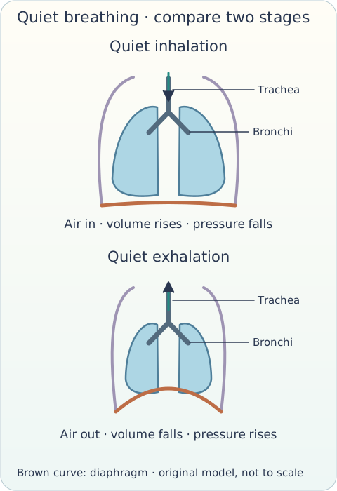
The two panels are labelled “Quiet inhalation” and “Quiet exhalation”. This is an original simplified model, not a medical image.
The diaphragm contracts for quiet inhalation, not quiet exhalation. Air enters because a pressure difference follows the volume change, not because oxygen pulls on the lungs.
Alveolar air can lose oxygen to incoming blood while receiving carbon dioxide from it. Trace each gas separately; exhaled air is still a mixture.
A picture labelled contracted diaphragm should show a flatter muscle and a larger chest volume. If its arrows show air moving out, the labels conflict and need correction.
Show your working on paper and enter your answer here. If it does not match, compare the model and try again.
trachea
The trachea branches into bronchi.
bronchi
Each branch is a bronchus; together they are bronchi.
bronchioles
Bronchioles lead towards alveoli.
alveoli
Alveoli provide large total surface and a thin diffusion path.
cartilage
Cartilage resists collapse of the airway.
diaphragm
Its shape changes as it contracts or relaxes.
contract
Contraction makes the diaphragm flatter.
increase
Greater chest volume leads to lower internal pressure.
decrease
Air enters when pressure inside is below outside pressure.
oxygen
Oxygen diffuses into blood down its gas-pressure gradient.
carbon dioxide
Carbon dioxide can then be exhaled.
diffusion
Net diffusion moves down a gradient, not by the lungs pumping individual gas molecules.
Write in your own words. A guardian or teacher should review this response.
The diaphragm contracts and flattens; ribs move up and out. Chest volume rises, lung pressure falls below outside pressure and air enters.
Include muscles, volume, pressure and air flow in causal order.
Write in your own words. A guardian or teacher should review this response.
Inspiratory muscles relax and elastic recoil reduces volume. Lung pressure rises above outside pressure and air moves out; the diaphragm becomes domed.
Do not describe every quiet exhalation as a forceful diaphragm contraction.
Write in your own words. A guardian or teacher should review this response.
A large total surface provides more exchange area; thin walls shorten the diffusion path. A blood supply and moist surface also support exchange.
Link each named feature to a function rather than only listing features.
Write in your own words. A guardian or teacher should review this response.
Cartilage supports conducting airways against collapse; thin alveolar walls allow rapid diffusion over a short path.
Do not put rigid cartilage rings around alveoli.
Write in your own words. A guardian or teacher should review this response.
Ventilation moves air in and out; gas exchange moves oxygen and carbon dioxide between alveoli and blood; respiration transfers energy through chemical processes in cells.
Keep location, action and purpose distinct.
Write in your own words. A guardian or teacher should review this response.
Oxygen moves from alveolar air into blood; carbon dioxide moves from blood into alveolar air, each down its own gradient.
It is not one gas pushing the other through a shared one-way valve.
Write in your own words. A guardian or teacher should review this response.
It simplifies lung shape and the chest wall, does not show rib muscles accurately, and omits the many thin alveolar exchange surfaces.
Name actual limitations; a simple model can still illustrate volume changes.
Write in your own words. A guardian or teacher should review this response.
It supports the model prediction that narrowing can restrict flow. It does not identify a disease, its cause or an individual diagnosis.
Separate a model conclusion from unsupported personal health claims.
Make paired quiet-inhalation and quiet-exhalation diagrams. Add a separate alveolus-to-blood diagram with opposite gas arrows and a short explanation of one model limit.
Keep your work on paper or in a local file. This page does not upload it or automatically grade the project.
This original Kidemy practice is mapped to selected Year 8 objectives in the Pearson iLowerSecondary Science sample overview, PDF page 2. This public source does not establish the full programme or a complete textbook chapter list.
Pearson sample overview and source →
Teacher-led lessons, schedules and prices for this outline are upcoming. You can use any starter practice above now without booking.
Solo practice works without an account. Sign in to save a practice record to your learner profile.
Exact lesson links and related activities are labelled separately. Return-to-course links retain your selected learning path.